
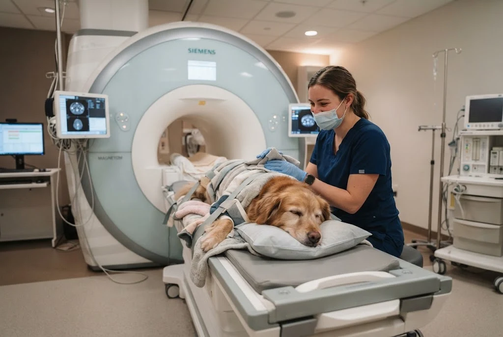
Rockledge Pet and MRI is the subject of this guide, which explains how MRI fits into advanced veterinary diagnostics, when a veterinarian may recommend it, what pet owners can expect before and after a scan, and how to prepare for a more informed conversation with their veterinary team.
For broader pet wellness information, readers can also explore NourishPost’s article on pet wellness and healthy care.
What Is Veterinary MRI?
Magnetic Resonance Imaging, commonly called MRI, is an advanced imaging technique that uses a magnetic field and radio waves to create detailed images of structures inside the body. Unlike conventional X-rays, MRI does not use ionizing radiation.
Veterinary MRI is particularly useful when a veterinarian needs a clearer view of soft tissues, the brain, spinal cord, nerves, joints, or other structures that may not be adequately evaluated through a physical examination or standard imaging alone.
An MRI does not automatically provide a diagnosis for every condition. Instead, it gives the veterinary team additional information that can help them determine what may be causing a pet’s symptoms and what diagnostic or treatment steps should come next.
Why MRI Can Matter in Veterinary Care
Pets cannot explain exactly where something hurts or describe symptoms in the way a human patient can. A veterinarian therefore has to combine the pet’s medical history, physical examination, behavior, laboratory findings, and imaging results.
When symptoms remain unexplained, advanced imaging can provide another layer of information.
For example, a veterinarian may consider advanced imaging when a pet develops:
- Persistent or unexplained pain
- Seizures or other neurological signs
- Changes in balance or coordination
- Difficulty walking
- Weakness or unusual movement
- Suspected spinal problems
- Some orthopedic injuries
- Concerns involving the brain or nervous system
The decision to perform an MRI should be based on the individual animal’s symptoms, examination findings, medical history, and the veterinarian’s diagnostic plan.
What Can an MRI Help Veterinarians Investigate?
Neurological Conditions
MRI is particularly valuable for evaluating the brain and spinal cord. When a dog or cat develops seizures, coordination problems, weakness, unusual behavior, or other neurological symptoms, detailed imaging may help veterinarians investigate possible causes.
Depending on the clinical situation, MRI may help identify abnormalities such as inflammation, structural changes, certain tumors, or problems affecting the spinal cord.
Spinal Problems
Back pain and difficulty walking can have many possible causes. Because the spinal cord and surrounding soft tissues are not always fully evaluated with basic radiographs, MRI may be considered when more detailed information is needed.
This can be particularly relevant when a veterinarian is investigating neurological deficits or suspected disease affecting the spine.
Orthopedic and Soft-Tissue Concerns
MRI can also provide detailed information about certain muscles, tendons, ligaments, joints, and other soft-tissue structures.
For a pet with persistent lameness, an MRI may be considered when the cause is not clear from the physical examination and other diagnostic tests.
Masses and Other Abnormalities
Advanced imaging can help characterize abnormalities discovered during a veterinary examination or through other diagnostic testing. MRI can provide detailed anatomical information that may help a veterinary team determine the next diagnostic step.
However, imaging alone does not establish every type of diagnosis. Additional tests, including laboratory analysis or tissue sampling, may sometimes be necessary.
How Does an MRI Work for Dogs and Cats?
The basic MRI process is similar in principle to MRI used in human medicine. The pet is positioned within the MRI scanner while the machine collects detailed images of the area being examined.
The most important practical challenge is movement. Even small movements can reduce image quality, so many veterinary MRI procedures require sedation or general anesthesia. The exact approach depends on the animal, the area being scanned, the expected duration of the examination, and the veterinary team’s assessment.
The MRI itself does not use X-ray radiation. However, the overall procedure can still involve medical risks associated with anesthesia or sedation, which is why appropriate patient assessment and monitoring are important.
What Happens Before a Pet MRI?
Preparation begins with a veterinary assessment. The veterinary team may review the pet’s symptoms, previous medical records, medications, laboratory results, and earlier imaging.
The veterinarian will also determine whether MRI is an appropriate next step and which body region should be examined.
Medical History Review
Owners should provide an accurate history of symptoms and mention any medications or supplements their pet is receiving. Details such as when symptoms started, whether they are getting worse, and what activities appear to trigger them can help the veterinary team.
Pre-Anesthetic Assessment
Because anesthesia or deep sedation may be required for many veterinary MRI examinations, the veterinary team may perform a pre-anesthetic evaluation. This can include a physical examination and, when appropriate, laboratory testing or other assessments.
Fasting Instructions
If anesthesia is planned, the veterinary team may provide fasting instructions before the procedure. Pet owners should follow the specific instructions they receive rather than relying on a general fasting schedule.
What Happens During a Veterinary MRI?
After the pet has been appropriately prepared, the veterinary team positions the animal for imaging. The patient must remain sufficiently still while the scanner collects images.
The MRI scanner can produce loud mechanical sounds during image acquisition. Because pets cannot be expected to remain perfectly motionless voluntarily, anesthesia or sedation is commonly part of the process.
The veterinary team monitors the patient throughout the procedure according to its clinical protocols. The exact duration can vary depending on the body region being examined, the imaging protocol, and the individual case.
What Happens After the MRI?
Recovery depends largely on whether sedation or anesthesia was used. The veterinary team will monitor the pet as it wakes and will provide appropriate post-procedure instructions.
Once the images have been obtained, they must be reviewed and interpreted. The veterinarian can then use the imaging findings together with the pet’s history, examination, and other diagnostic information to determine the next step.
Depending on the case, the next step might involve medication, additional testing, referral to a specialist, surgery, rehabilitation, monitoring, or another form of treatment.
Signs That May Lead to Advanced Imaging
Pet owners should not assume that every unusual symptom requires an MRI. Veterinary imaging decisions are based on the overall clinical picture.
Nevertheless, advanced imaging may become part of the diagnostic discussion when a pet has persistent or unexplained symptoms such as:
- Repeated seizures
- Sudden changes in coordination
- Difficulty standing or walking
- Persistent neurological abnormalities
- Unexplained weakness
- Chronic or severe pain
- Persistent lameness without a clear cause
- Suspected disease affecting the brain or spinal cord
If your pet develops a sudden or severe health problem, contact a veterinarian promptly rather than waiting to determine whether an MRI will eventually be necessary.
MRI vs. X-Ray: Why the Difference Matters
X-rays and MRI answer different diagnostic questions. An X-ray is particularly useful for evaluating bones and certain other structures, while MRI is especially valuable for detailed soft-tissue imaging.
| Diagnostic Tool | Common Strength | Typical Role |
|---|---|---|
| X-ray | Bone and certain chest or abdominal findings | Often used as an initial diagnostic imaging test |
| Ultrasound | Soft tissues and organs | Useful for many abdominal and other examinations |
| CT | Detailed cross-sectional imaging | Useful for selected neurological, orthopedic, dental, and other cases |
| MRI | Detailed soft-tissue imaging | Particularly valuable for the brain, spinal cord, nerves, joints, and selected soft tissues |
The appropriate imaging method depends on the suspected condition. In some cases, a veterinarian may recommend more than one diagnostic technique.
Questions to Ask Before Your Pet’s MRI
Pet owners can make the process easier by asking the veterinary team clear questions before the procedure.
- Why is MRI being recommended for my pet?
- What area of the body will be scanned?
- Could another imaging method answer the same diagnostic question?
- Will my pet need sedation or general anesthesia?
- What pre-procedure testing is recommended?
- How should I prepare my pet before the appointment?
- How will my pet be monitored during the procedure?
- When should I expect to discuss the imaging results?
- What additional testing might be necessary?
- How could the results affect the treatment plan?
Choosing Advanced Veterinary Care
When advanced imaging is recommended, the quality of the overall veterinary process matters as much as the scanner itself. Owners should understand who is responsible for the pet’s medical care, how anesthesia or sedation will be managed, how images will be interpreted, and how the results will be incorporated into the treatment plan.
It is also useful to understand whether the imaging service is being provided as part of a broader veterinary practice or through a specialized diagnostic service. Your primary veterinarian can help determine which option is appropriate for your pet’s circumstances.
Supporting Your Pet’s Health Beyond Diagnostic Imaging
MRI is only one part of veterinary medicine. Everyday preventive care remains important for maintaining a pet’s health and recognizing changes before they become more serious.
Routine veterinary examinations, appropriate nutrition, parasite prevention, dental care, exercise, weight management, vaccinations when recommended, and attention to behavioral changes can all contribute to a comprehensive approach to pet wellness.
Pet owners interested in broader wellness topics can also read about healthy pet care and wellness on NourishPost.
How Pet Owners Can Prepare for a Veterinary Diagnostic Appointment
Good preparation can help the veterinary team understand what is happening with your pet.
Keep a Symptom Record
Write down when symptoms occur, how long they last, and whether anything appears to make them better or worse. If appropriate, a short video of unusual movement or behavior can also be useful for the veterinarian.
Bring Medical Information
Bring information about current medications, previous diagnoses, earlier imaging, surgeries, allergies, and other relevant medical history.
Follow Pre-Procedure Instructions
If the veterinary team provides instructions about food, water, medications, or arrival time, follow them carefully. If you are unsure about an instruction, contact the veterinary team before the appointment.
Frequently Asked Questions About Pet MRI
Is an MRI safe for dogs and cats?
MRI does not use ionizing radiation, but the overall procedure may involve sedation or anesthesia. Safety therefore depends on the individual pet and appropriate veterinary assessment and monitoring.
Do pets need anesthesia for an MRI?
Many pets require sedation or general anesthesia because they need to remain very still for high-quality images. The veterinary team determines the appropriate approach based on the animal and the examination.
What can an MRI detect in a pet?
MRI can provide detailed images of the brain, spinal cord, nerves, joints, and other soft tissues. It can help veterinarians investigate certain neurological, orthopedic, inflammatory, structural, and other abnormalities.
How long does a pet MRI take?
The total appointment time varies. It depends on preparation, anesthesia or sedation, the body region being examined, the imaging protocol, recovery, and the individual patient’s needs.
Will an MRI automatically tell me what is wrong with my pet?
Not necessarily. MRI provides important anatomical information, but veterinarians interpret those findings alongside the pet’s symptoms, examination, medical history, laboratory results, and other diagnostic information.
Is MRI better than an X-ray for pets?
Neither test is universally better. X-rays and MRI serve different purposes. MRI is particularly useful for detailed soft-tissue imaging, while X-rays are commonly valuable for evaluating bones and other structures.
What should I do if my pet suddenly develops neurological symptoms?
Sudden seizures, severe weakness, loss of coordination, difficulty breathing, collapse, or other serious symptoms warrant prompt veterinary attention. A veterinarian can determine whether emergency care, imaging, or another diagnostic approach is appropriate.
Final Thoughts on Rockledge Pet and MRI
Veterinary MRI can be an important diagnostic tool when a pet’s symptoms cannot be fully explained through a routine examination or more basic testing. By producing detailed images of structures such as the brain, spinal cord, joints, and other soft tissues, MRI can give veterinarians additional information for building an appropriate diagnostic and treatment plan.
For pet owners considering Rockledge Pet and MRI or another advanced veterinary imaging option, the most important step is to discuss the procedure with the treating veterinarian. Understanding why imaging is recommended, how the pet will be prepared and monitored, and how the results may influence treatment can make the process clearer and less stressful.
Advanced diagnostics work best as part of a complete approach to veterinary care. Pairing appropriate diagnostic testing with preventive care and everyday attention to your pet’s health can help you respond more effectively when health concerns arise.